2015年比較腫瘍学常陸宮賞受賞者
The Awardee of the 2015 Prince Hitachi Prize for Comparative Oncology
"Elucidation of the General Principles of Vertebrate Organogenesis Using Small Fish"
小型魚類を用いた脊椎動物の器官形成原理の解明
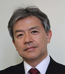 Hiroyuki Takeda
Professor, Graduate School of Science University of Tokyo
武田 洋幸 博士
東京大学大学院理学系研究科 教授
略歴
武田洋幸博士は、1982年3月に東京大学理学部動物学教室を卒業し、同年4月に同大学院理学系研究科動物学専門課程修士課程に進学、水野丈夫教授のもとで、マウスとラットを用いて哺乳類前立腺発生と性ホルモンの研究に従事、1987年5月に、「哺乳類前立腺発生におけるアンドロゲン依存性組織間相互作用のステロイドオートラジオグラフィー法による解析」の研究で博士(理学)を授与された。助手在職中の1986年と1989年に、それぞれ10か月間英国ストレンジウェィ研究所、1年間シカゴ大学に滞在して、前立腺がんの研究を行った後、1990年4月に理化学研究所ライフサイエンス筑波研究センターの研究員となり、天沼宏博士のもとでマウス白血病ウイルスによる白血病発症の研究に従事した。
一方、1991年からゼブラフィッシュを用いた発生学の研究を独自に開始した。1993年11月には名古屋大学理学部助教授(黒岩 厚研究室)、1999年3月には国立遺伝学研究所教授(初期発生部門)、2000年4月からは現職の東京大学大学院理学系研究科教授となり、この間一貫して小型魚類の発生遺伝学、ゲノム生物学の研究を推進している。2008年には「小型魚類を用いた脊椎動物発生機構の普遍的メカニズムの解明」で日本動物学会賞を受賞し、2010年から2014年度新学術領域「秩序形成ロジック」領域代表、2013年から科学技術振興機構創造的研究推進事業(CREST)・研究領域「生命動態の理解と制御のための基盤技術の創出」で、研究課題「DNA3次元クロマチン動態の理解と予測」の研究代表者を務めている。また2014年から日本動物学会会長に就任している。
Personal History Outline
Dr. Hiroyuki Takeda was born on 17 October 1958 in Niigata City, Japan. He graduated from the Department of Biological Sciences, Faculty of Science, University of Tokyo in 1982 and started his study on hormone-dependent tissue interaction during the development of the rodent prostate gland, under the supervision of Professor Takeo Mizuno. In 1987, he was granted a Doctor of Science under the thesis entitled "Steroid autoradiographic analysis of androgen-dependent tissue interactions in the development of mammalian prostate glands". He also worked on prostate cancer in the Strangeways Research Laboratory (Cambridge, UK) with Dr. Ilse Lasnitzki for 10 months (1986-1987) and in the Urological Department of Chicago University with Dr. Chawnshang Chang for 1 year (1989-1990) as a visiting scholar. He returned to Japan in 1990 to work as a Research Scientist at RIKEN where he first studied virus-induced leukemia in mouse in the laboratory of Dr. Hiroshi Amanuma.
During this period, he introduced the zebrafish system to his own project on axis formation and organogenesis in vertebrates. In 1994, he joined the laboratory of Professor Atsushi Kuroiwa as an Associate Professor of the Faculty of Science, University of Nagoya, and continued his zebrafish work. After 5 years in Nagoya, he moved to the National Institute of Genetics, Mishima, to take a position of Professor of the Division of Early Embryogenesis in 1999. There he started using medaka in addition to zebrafish.
In 2000, Dr. Takeda came back to his original laboratory and was appointed as a professor at the University of Tokyo. In 2008, he was awarded the Zoological Society Prize by the Zoological Society of Japan under the title "Analysis of Early Embryogenesis using small fish as a model". Between 2010 and 2014, he served as the head investigator of the Grant-in-Aid for Scientific Research on Innovative Areas entitled by “From Molecules and Cells to Organs: Trans-Hierarchical Logic for Higher-Order Pattern and Structures, granted by the Ministry of Education, Culture, Sports, Science and Technology Japan,”. At present, he is also serving as the principal investigator of the Core Research Project for Evolutional Science and Technology (CREST) entitled "Understanding and Predicting 3D Chromatin Dynamics (2013-2018)", granted by the Japan Science and Technology Corporation (JST),. Since 2014, he has been appointed the President of the Zoological Society of Japan.
研究業績
武田洋幸博士は、1991年に独自に我が国ではじめてゼブラフィッシュを脊椎動物の発生研究やヒト疾患モデルとして導入した。当時この魚は、短い世代交代時間、多産、透明な胚などの特徴を有し、また比較的単純な体制にもかかわらずヒトと同じような発生メカニズムを持っている優れたモデル動物として認識され始めていた。博士は2000年には、日本発のモデル実験系であるメダカも研究に取り入れ、以来これら小型魚類の化学発癌剤ENU誘発の突然変異体、ならびに自然突然変異体を利用して、脊椎動物の普遍的な発生原理に関して、世界的な業績を着実に上げ続けてきた。
武田博士が得た最初の特筆すべき成果は、魚類ではじめてとなる中胚葉誘導組織の発見である(1996年)。さらに、近藤滋博士(大阪大学)との共同研究による、数理生物学とゼブラフィッシュの実験発生学を融合した「分節時計の作動原理」の解明が重要である。体節形成は脊椎動物の体幹部の分節性を規定する重要なイベントであるが、体節は、神経管の両脇にある中胚葉細胞塊が前方から後方へ、動物固有の一定のリズム(ヒト6時間程度、マウス2時間、ニワトリ90分、ゼブラフィッシュ30分など)で一対ずつ形成される。武田博士は、体節形成に関係する遺伝子のうち、重要な転写因子Tbx24の機能解析とFgf/MAPKシグナルの作用機序の解明を行い(2000-2002年)次に「分節時計」の研究に焦点をあてた研究を開始した。体節を形成する組織は数千から数万個の時計細胞の集合体である。個々の時計細胞内では、ヘアリと呼ばれる抑制性転写因子の発現量が周期的に変動することを利用して時を刻む。このON/OFFのリズムが細胞ごとにバラバラのままだと体節を作れなくなり、脊椎骨融合症となる。体節形成における時計細胞の同調性の原因についてはこれまで不明であったが、武田博士らは、この分節時計の基本的な作動原理を、変異体、細胞の移植、数学的シミュレーションを駆使して解明した。その結果、分節時計は、最小単位である時計細胞が、ノッチ(受容体)・デルタ(リガンド)という細胞表面で機能するタンパク質を使って互いに手を取り合うことで一つの集団をつくり、同調して振動することが明らかとなった。さらに詳細な観察の結果、正常に作動している分節時計であっても、高頻度で起こる細胞分裂などの影響で、時計細胞間の転写タイミングの“ゆらぎ”(ノイズ)が生じるが、これらのノイズもまた、ノッチ-デルタを介した位相同期機構により効率よく軽減されることも見出した(2006年)。すなわち、体節時計の作動原理は蛍の発光同期に見られる結合振動系と同じ原理であった。この研究は、組織を作る細胞間の遺伝子プログラムの安定性を保証するしくみを示しており、細胞から構成される組織が統一体として機能するという現象の根幹にふれる重要なものである.本研究は、統一性のある組織からの逸脱を特徴とするがん細胞の本態解明にも将来貢献すると期待される。
武田博士はさらに、メダカ変異体を用いてヒトの病気の原因解明につながる研究を行っている。その一つが、2000から2002年に国立遺伝学研究所で行われたがん原化学物質ENU誘発の変異体スクリーニングで得られた、心臓ループの逆転を起こす突然変異体ktuの研究である。この変異体は嚢胞腎も発症する。解析の結果、ktuはダイニン腕の細胞質中でのアッセンブリーに必須の新規の遺伝子で、ダイニン腕形成を通して繊毛の運動性を支配しており、繊毛の異常で起こるカルタゲナー症候群(気管支拡張、慢性の副鼻腔炎、内臓逆位、男性不妊など)や、多発性嚢胞腎、多指症など、ヒト繊毛病(ciliopathy)の原因遺伝子の一つであった(Heymut Omran博士、Freiburg UniversityおよびDavid R. Mitchell博士、UNY Upstate Medical Universityとの共同研究)。このktu遺伝子は脊椎動物だけでなく、昆虫類、さらにクラミドモナスにも存在し、その変異が脊椎動物同様に鞭毛の運動異常を引き起こすことが分かった(2008年)。このように、ktuは、原生生物からヒトに至るまで、進化的に保存されている普遍的な遺伝子であり、繊毛システムを介する生命現象(細胞周期を含む)に広く関与していた。メダカ変異体を用いてヒトの繊毛病の原因遺伝子を明らかにしたこの重要な業績は、発生生物学および病理学と同時に比較腫瘍学の収穫である。
Academic Achievement
Dr. Hiroyuki Takeda introduced zebrafish for his research by himself in 1991 while at RIKEN for the first time in Japan in order to study vertebrate development and human genetic diseases. Zebrafish emerged as a model organism,characterized by its short generation time, a large number of offspring and transparency of embryos. The development of the zebrafish is very similar to that of higher vertebrates, including humans. In 2000, he started using medaka, a Japanese killifish, in addition to the zebrafish. The medaka fish is a Japanese-made vertebrate model with the unique features of a smaller genome and various established inbred lines suitable for genetics and genomics. Over the past two decades, his works with ENU (a chemical mutagen)-induced and spontaneous fish mutants, have greatly contributed to the understanding of the general principles of vertebrate organogenesis.
Dr. Takeda’s first well-known study in his zebrafish research was the identification of a yolk cell as a mesoderm inducer in teleosts. A more important series of works followed in the next ten years, which led to the elucidation of the basic principle of somite segmentation. Somites are formed one by one from the anterior end of the un-segmented mesoderm (presomitic mesoderm, PSM) in an anterior-to-posterior direction, which is a crucial event in the creation of a metameric body pattern in vertebrates. One of the most important aspects of somitogenesis is its strict periodicity; a pair of somites is formed every 30 min for zebrafish, 90 min for chickens, 120 min for mice and 6 hours for humans. Because of this, it had been postulated that a segmentation clock exists in the PSM, creating this periodicity during somite formation. If segmentation goes wrong, humans suffer from synostosis of the spine. After having analyzed the essential roles of two transcription factors, Mesp and Tbx24, in somite segmentation, and how these factors are regulated by Fgf/MAPK signaling in the PSM, he focused on the segmentation clock, in particular, how numerous cells behave like a single clock. The segmentation clock consists of numerous cellular oscillators which are driven by a negative-feedback loop of Hairy transcription factor. They are linked through Notch-dependent intercellular coupling and display the synchronous expression of clock genes. Combining transplantation experiments in zebrafish with mathematical simulations (in collaboration with Dr. Shigeru Kondo, Osaka University), he demonstrated that the segmentation clock behaves as a “coupled oscillators,” a mechanism that also underlies the synchronous flashing seen in fireflies (2006). With this mechanism the segmentation clock forms a robust system which is resistant to the effects of developmental noise such as stochastic gene expression and active cell proliferation. This work highlights the presence of higher-order mechanisms that ensure the stability of the genetic program for precise pattern formation in multicellular organisms. Since the caner cell is characterized by its tendency to break away from harmonized normal tissue, Dr Takeda’s work may contribute to enabling a deeper insight into the nature of cancer cells in the future.
Between 2000 and 2002, Dr. Takeda conducted an ENU-mutagenesis screening of medaka at the National Institute of Genetics and isolated various developmental mutants, some of which served as human disease models. One such mutant is ktu, originally isolated by reverse heart-looping, which turns out to be caused by defects in ciliary motility. Dysfunction of the cilium is the basis for multiple human genetic disorders that have collectively been called ‘ciliopathy’ and whose phenotypes are quite diverse, e.g. Kartagener syndrome (situs inversus, male sterility, recurrent respiratory tract infections), polydactyly, and polycystic kidney. Indeed the ktu also develops polycystic kidney disease. His research group demonstrated that the ktu gene encodes a novel cytoplasmic protein conserved from ciliated unicellular organisms to higher mammals and required for cytoplasmic pre-assembly of axonemal dyneins. Furthermore, mutations were identified in the homologous gene of two human ciliopathy families and Chlamydomonas mutant, pf13 (in collaboration with Dr. Heymut Omuran, Freiburg University and Dr. David R. Mitchell, SUNY Upstate Medical University, respectively). Taken together, starting from a tiny medaka mutant, his group identified a long-neglected gene which is universally required for all ciliated organisms and participates in a variety of biological phenomena through regulating ciliary motility (2008). This is an important contribution to the developmental biology, the pathology and also to the comparative oncology.