The Awardee of the 2015 Prince Hitachi Prize for Comparative Oncology
"Elucidation of the General Principles of Vertebrate Organogenesis Using Small Fish"
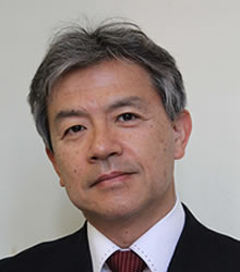 Hiroyuki Takeda
Professor, Graduate School of Science University of Tokyo
Personal History Outline
Dr. Hiroyuki Takeda was born on 17 October 1958 in Niigata City, Japan. He graduated from the Department of Biological Sciences, Faculty of Science, University of Tokyo in 1982 and started his study on hormone-dependent tissue interaction during the development of the rodent prostate gland, under the supervision of Professor Takeo Mizuno. In 1987, he was granted a Doctor of Science under the thesis entitled "Steroid autoradiographic analysis of androgen-dependent tissue interactions in the development of mammalian prostate glands". He also worked on prostate cancer in the Strangeways Research Laboratory (Cambridge, UK) with Dr. Ilse Lasnitzki for 10 months (1986-1987) and in the Urological Department of Chicago University with Dr. Chawnshang Chang for 1 year (1989-1990) as a visiting scholar. He returned to Japan in 1990 to work as a Research Scientist at RIKEN where he first studied virus-induced leukemia in mouse in the laboratory of Dr. Hiroshi Amanuma.
During this period, he introduced the zebrafish system to his own project on axis formation and organogenesis in vertebrates. In 1994, he joined the laboratory of Professor Atsushi Kuroiwa as an Associate Professor of the Faculty of Science, University of Nagoya, and continued his zebrafish work. After 5 years in Nagoya, he moved to the National Institute of Genetics, Mishima, to take a position of Professor of the Division of Early Embryogenesis in 1999. There he started using medaka in addition to zebrafish.
In 2000, Dr. Takeda came back to his original laboratory and was appointed as a professor at the University of Tokyo. In 2008, he was awarded the Zoological Society Prize by the Zoological Society of Japan under the title "Analysis of Early Embryogenesis using small fish as a model". Between 2010 and 2014, he served as the head investigator of the Grant-in-Aid for Scientific Research on Innovative Areas entitled by “From Molecules and Cells to Organs: Trans-Hierarchical Logic for Higher-Order Pattern and Structures, granted by the Ministry of Education, Culture, Sports, Science and Technology Japan,”. At present, he is also serving as the principal investigator of the Core Research Project for Evolutional Science and Technology (CREST) entitled "Understanding and Predicting 3D Chromatin Dynamics (2013-2018)", granted by the Japan Science and Technology Corporation (JST),. Since 2014, he has been appointed the President of the Zoological Society of Japan.
Academic Achievement
Dr. Hiroyuki Takeda introduced zebrafish for his research by himself in 1991 while at RIKEN for the first time in Japan in order to study vertebrate development and human genetic diseases. Zebrafish emerged as a model organism,characterized by its short generation time, a large number of offspring and transparency of embryos. The development of the zebrafish is very similar to that of higher vertebrates, including humans. In 2000, he started using medaka, a Japanese killifish, in addition to the zebrafish. The medaka fish is a Japanese-made vertebrate model with the unique features of a smaller genome and various established inbred lines suitable for genetics and genomics. Over the past two decades, his works with ENU (a chemical mutagen)-induced and spontaneous fish mutants, have greatly contributed to the understanding of the general principles of vertebrate organogenesis.
Dr. Takeda’s first well-known study in his zebrafish research was the identification of a yolk cell as a mesoderm inducer in teleosts. A more important series of works followed in the next ten years, which led to the elucidation of the basic principle of somite segmentation. Somites are formed one by one from the anterior end of the un-segmented mesoderm (presomitic mesoderm, PSM) in an anterior-to-posterior direction, which is a crucial event in the creation of a metameric body pattern in vertebrates. One of the most important aspects of somitogenesis is its strict periodicity; a pair of somites is formed every 30 min for zebrafish, 90 min for chickens, 120 min for mice and 6 hours for humans. Because of this, it had been postulated that a segmentation clock exists in the PSM, creating this periodicity during somite formation. If segmentation goes wrong, humans suffer from synostosis of the spine. After having analyzed the essential roles of two transcription factors, Mesp and Tbx24, in somite segmentation, and how these factors are regulated by Fgf/MAPK signaling in the PSM, he focused on the segmentation clock, in particular, how numerous cells behave like a single clock. The segmentation clock consists of numerous cellular oscillators which are driven by a negative-feedback loop of Hairy transcription factor. They are linked through Notch-dependent intercellular coupling and display the synchronous expression of clock genes. Combining transplantation experiments in zebrafish with mathematical simulations (in collaboration with Dr. Shigeru Kondo, Osaka University), he demonstrated that the segmentation clock behaves as a “coupled oscillators,” a mechanism that also underlies the synchronous flashing seen in fireflies (2006). With this mechanism the segmentation clock forms a robust system which is resistant to the effects of developmental noise such as stochastic gene expression and active cell proliferation. This work highlights the presence of higher-order mechanisms that ensure the stability of the genetic program for precise pattern formation in multicellular organisms. Since the caner cell is characterized by its tendency to break away from harmonized normal tissue, Dr Takeda’s work may contribute to enabling a deeper insight into the nature of cancer cells in the future.
Between 2000 and 2002, Dr. Takeda conducted an ENU-mutagenesis screening of medaka at the National Institute of Genetics and isolated various developmental mutants, some of which served as human disease models. One such mutant is ktu, originally isolated by reverse heart-looping, which turns out to be caused by defects in ciliary motility. Dysfunction of the cilium is the basis for multiple human genetic disorders that have collectively been called ‘ciliopathy’ and whose phenotypes are quite diverse, e.g. Kartagener syndrome (situs inversus, male sterility, recurrent respiratory tract infections), polydactyly, and polycystic kidney. Indeed the ktu also develops polycystic kidney disease. His research group demonstrated that the ktu gene encodes a novel cytoplasmic protein conserved from ciliated unicellular organisms to higher mammals and required for cytoplasmic pre-assembly of axonemal dyneins. Furthermore, mutations were identified in the homologous gene of two human ciliopathy families and Chlamydomonas mutant, pf13 (in collaboration with Dr. Heymut Omuran, Freiburg University and Dr. David R. Mitchell, SUNY Upstate Medical University, respectively). Taken together, starting from a tiny medaka mutant, his group identified a long-neglected gene which is universally required for all ciliated organisms and participates in a variety of biological phenomena through regulating ciliary motility (2008). This is an important contribution to the developmental biology, the pathology and also to the comparative oncology.