
![]()
- HOME
- Laboratories
- Clinical Chemotherapy
- Projects

Research introduction and trait
In our laboratory, we conduct the study on various types of cancer. We accomplish the study about the molecular biochemical character, the susceptibility for the molecular target drugs, and the resistant mechanism with original idea in a wide variety of types of cancer, such as malignant lymphoma, acute leukemia, breast cancer, bone metastases, gastric cancer, prostate cancer, and renal cell cancer. A particularly distinguishing is the collaborative investigation with the pathologists, and we takes pride in studying for the benefit of patients by using clinical specimen obtained from patients in the clinical department. We regard the development of the field of new treatment of the cancer as a mission, by elucidating the susceptibility and resistant mechanism for the molecular target drugs in each type of cancer, and by developing inhibitor and the activator for these molecules target.
Projects
- The significance of circulating tumor cells and circulating endothelial cells in solid tumor
- The clinical trial-based translational researches including breast cancer, bone-metastatic cancer and caner of unknown primary
- Prediction of therapy effect of molecular target drugs and mutation analysis of the target molecules
- sThe application of fluorescent imaging to the search of a biomarkers predicting the therapeutic effects
- The application of fluorescent imaging to the diagnosis of hematopoietic tumors (fluorescence 3D blood atlas)
- The functional analysis of novel protein RNF137 involved in antitumor effect of imatinib
- The biomarker study in the gastrointestinal carcinoma chemotherapy
- Discovery of novel biomarker and molecular target therapy, based on the carcinogenic mechanism in the urologic cancer
The application of fluorescent imaging to the search of a biomarkers predicting the therapeutic effects
OLYMPUS-Bio-Imaging lab was established by the collaboration of JFCR and OLYMPUS corporation. We aim to establish the novel imaging techniques that will be applied to the cancer screening, made-to-order therapies or translational research. We are trying to develop the strategy to predict practical clinical consequence of molecule target drugs, and are evaluating clinic cases of cancer in ex vivo fluorescent imaging.
The application of fluorescent imaging to the search of the biomarkers predicting the therapeutic effects
In recent years, therapeutic antibodies have greatly improved the efficacy of chemotherapy regimens. On the other hand, it becomes critical to stratify the patient expected to acquire a good treatment outcome because treatment costs of antibody therapy are exceedingly large. So, we evaluate the susceptibility of the tumor cells derived from patient who received antibody therapy by the procedure using live-cell-imaging, and try to develop the methods that can predict the outcome of the antibody therapy by cooperation of the many patients of the Cancer Institute Hospital.
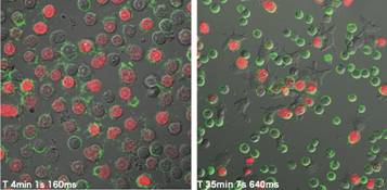
[Figure legend]Imaging of complement-dependent cell cytotoxicity and antibody-dependent cell-mediated cytotoxicity by a CD20 antibody
These are the examples observed the manner that tumor cells from a patient of malignant lymphoma were killed by anti-CD20 monoclonal antibody drug. Fluorescent-labeled antibody drug (ritxuimab-Alexa488) bound to the tumor cells and then killed them by complement factors (left) or natural killer cells (right). [green: rituximab, red: Propidium iodide, by OLYMPUS FV-1000 confocal microscope]
The application of fluorescent imaging to the diagnosis of hematopoietic tumors (fluorescence 3D blood atlas
As cell surface proteins, nucleus, and organelles can be labeled with fluorescent agents without severe damaging to the cells by the advances of fluoro-labeling technology, we can acquire various information from living cells. We apply the multi-color fluorescent imaging to the living biopsy of hemopoietic malignant cells and aim at the application to rapid diagnosis by observing cells in three-dimensional assessment.
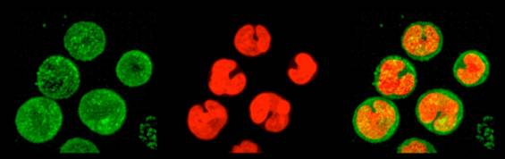
[Figure legend] An analysis case of NK leukemic cells
The peripheral blood mononuclear cells derived from a patient with suspected leukemia with CD56 antigen and nucleus without fixing. [green: CD56, red: nucleus, by OLYMPUS FV-1000 confocal microscope]
![]()